
CT vs MRI
in Veterinary Medicine
Technical and Diagnostic Differences Between MRI and CT in Veterinary Medicine
Magnetic Resonance Imaging (MRI) and Computed Tomography (CT) are advanced imaging modalities widely used in veterinary medicine. While both provide cross-sectional images of the body, they differ significantly in their underlying technology, image acquisition methods, and clinical applications. Understanding these differences is essential for selecting the most appropriate modality based on the patient’s condition and diagnostic needs. A detailed technical analysis follows at the bottom of this page. If you prefer to see this in video form, please view our CT vs MRI video.
Technical Differences
Imaging Physics
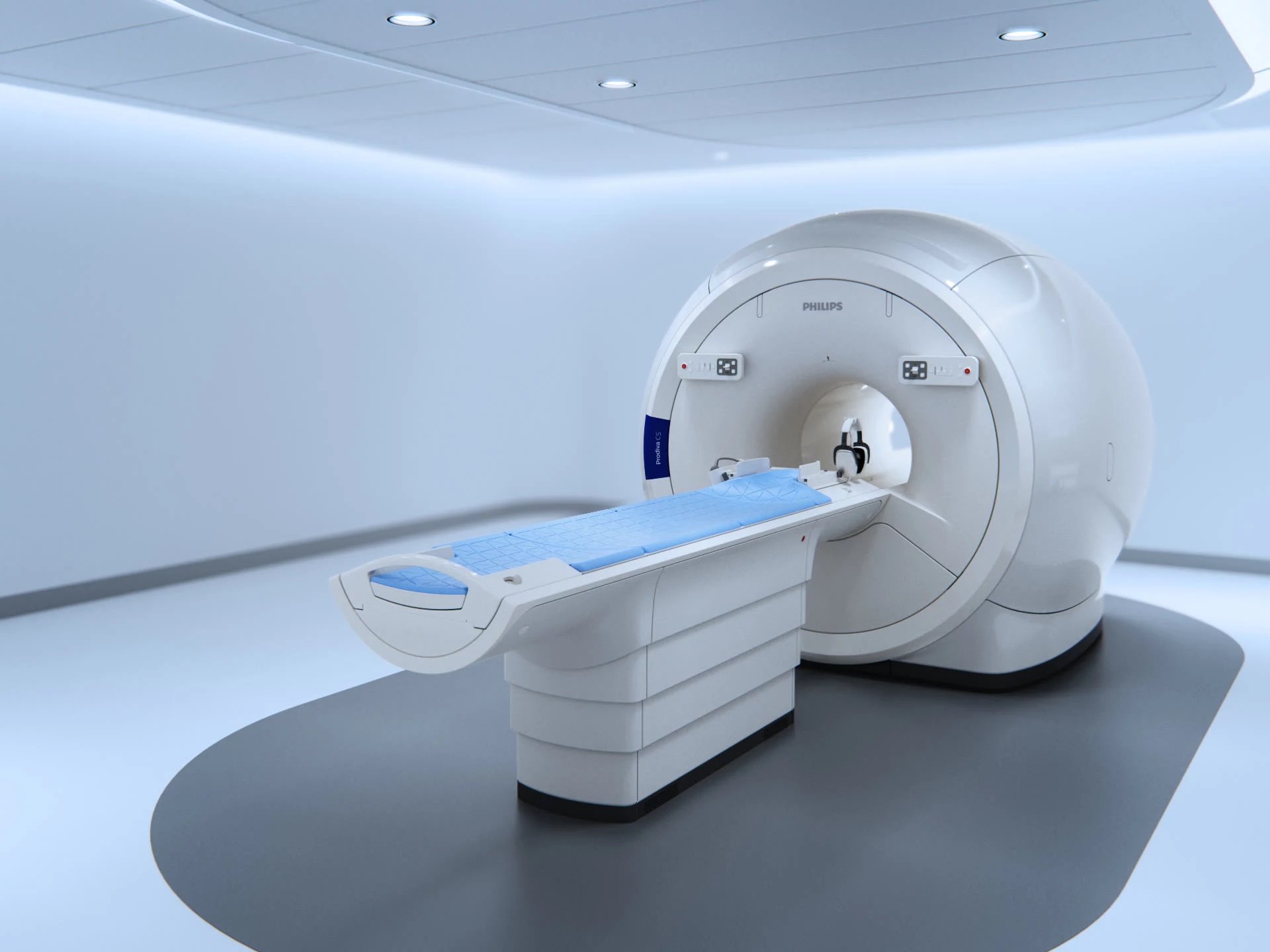
MRI: Utilizes strong magnetic fields and radiofrequency pulses to manipulate hydrogen nuclei within tissues, measuring their response to generate images.
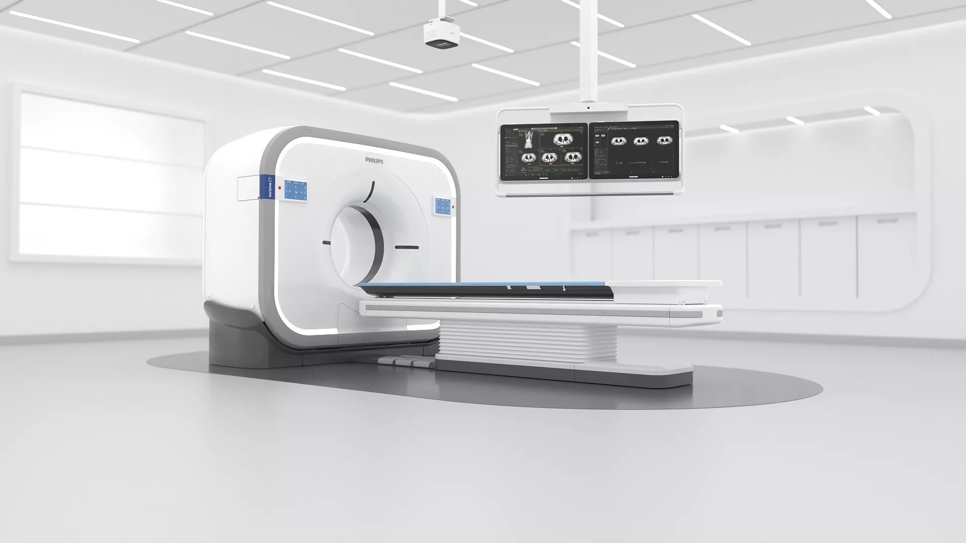
CT: Uses X-ray beams and computer processing to measure tissue density differences, producing grayscale cross-sectional images.
Tissue Contrast and Resolution
MRI: Provides excellent soft tissue contrast, distinguishing between subtle variations in tissue composition, such as gray and white matter in the brain.
CT: Offers high spatial resolution, particularly for bony structures, allowing precise visualization of fractures and mineralized tissue.
Scan Time and Patient Considerations
MRI: Requires longer scan times (30–90 minutes) and is highly sensitive to motion, necessitating general anesthesia in most veterinary patients.
CT: Provides rapid image acquisition (seconds to minutes), often requiring only sedation, though anesthesia may still be needed for optimal image quality.
Radiation Exposure
MRI: Does not use ionizing radiation, making it safer for repeated imaging.
CT: Involves ionizing radiation, requiring adherence to safety protocols to minimize exposure.
Artifacts and Image Limitations
MRI: Susceptible to motion artifacts, metal interference, and flow-related distortions.
CT: Can exhibit beam hardening artifacts, motion artifacts, and partial volume effects that may impact image interpretation.
Diagnostic Applications and Key Indications
Neurological Imaging
MRI is the modality of choice for brain and spinal cord evaluation due to its superior soft tissue contrast.
Common indications include:
Seizures, brain tumors, encephalitis, and infarcts
Intervertebral disc disease (IVDD) and spinal cord compression
Myelitis, meningitis, and demyelinating diseases
Musculoskeletal and Orthopedic Imaging
MRI is best for soft tissue structures such as tendons, ligaments, and muscles.
CT is preferred for bony abnormalities and fractures.
Indications include:
MRI: Tendon and ligament injuries, joint capsule damage, and muscle atrophy
CT: Complex fractures, osteochondrosis, and bone tumors
Thoracic and Pulmonary Imaging
CT is superior for lung and thoracic imaging due to its ability to detect subtle lung pathology.
Common indications include:
Pulmonary masses, metastases, and interstitial lung disease
Thoracic trauma and rib fractures
Pleural effusion characterization
Nasal and Sinus Disease
CT is the preferred modality for evaluating nasal passages, sinuses, and skull structures.
Indications include:
Chronic rhinitis, fungal infections, and neoplasia
Foreign body detection
Bony destruction or remodeling
Middle and Inner Ear Disease
CT is better for bony involvement, while MRI is superior for soft tissue and nerve evaluation.
Indications include:
Otitis media/interna
Tympanic bulla involvement
Vestibular dysfunction of central vs. peripheral origin
Abdominal and Oncologic Imaging
CT is widely used for abdominal mass evaluation and oncologic staging.
Indications include:
Detection of metastases, lymph node involvement, and vascular invasion
Evaluation of organ masses, mineralized lesions, or contrast-enhanced abnormalities
Whole-body imaging for cancer staging
Vascular and Cardiac Imaging
CT angiography is the preferred method for vascular evaluation.
Indications include:
Congenital vascular anomalies (e.g., portosystemic shunts)
Cardiac and great vessel abnormalities
Pre-surgical planning for vascular tumors
Summary of Clinical Use Cases
MRI is best for soft tissue imaging, particularly for neurological conditions, tendon and ligament injuries, and soft tissue tumors.
CT is best for bone, lung, and vascular imaging, making it ideal for fractures, nasal disease, pulmonary metastases, and oncologic staging.
Both modalities are complementary, and the choice of imaging depends on the clinical question, patient stability, and specific diagnostic needs. Understanding the strengths and limitations of each technique ensures optimal patient care and accurate diagnosis.
Comparison of Diagnostic and Technical Factors Between MRI and CT in Veterinary Medicine
Diagnostic Factors

Technical Factors
